Editor's Note, Volume 6 Issue 4

D’Arcy Little, MD, CCFP, FRCPC
Medical Director, JCCC and HealthPlexus.NET

D’Arcy Little, MD, CCFP, FRCPC
Medical Director, JCCC and HealthPlexus.NET


Physicians usually become adept at choosing medications for the complaints and illnesses that patients bring to their attention.
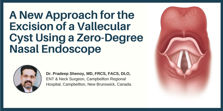
Dr. Pradeep Shenoy, MD, FRCS, FACS, DLO,
ENT & Neck Surgeon, Campbellton Regional Hospital, Campbellton, New Brunswick, Canada.
| Abstract: This case study reviews the clinical presentation and treatment of a patient’s vascular cyst. Though it is a rare diagnosis/condition, it could be a medical emergency for an individual of any age. Described here is a new approach for the complete excision of the vallecular cyst. |
| Key Words: vallecular cyst, excision, zero-degree nasal endoscope |
| Vallecular cysts are retention cysts in the Vallecular—a space between the base of the tongue, epiglottis and lateral pharyngeal wall. |
| They are triggered by acid reflux and smoking. |
| Vallecular cyst can be seen in CT Scan and laryngoscopy examination. |
| The access is difficult trans-orally. Here we are describing New approach using a zero-degree endoscope. |
| Vallecular cysts are rare in the paediatric and adult age groups. |
| Vallecular cysts can present as asymptomatic when small, however, when big they can present as a feeling of some food stuck in the throat or pain. |
| In emergency situation can block the food and airway passage and require emergency treatment. |
| To have access to full article that these tools were developed for, please subscribe. The cost to subscribe is $80 USD per year and you will gain full access to all the premium content on www.healthplexus.net, an educational portal, that hosts 1000s of clinical reviews, case studies, educational visual aids and more as well as within the mobile app. |
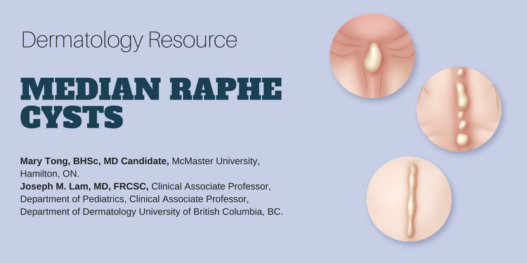
Mary Tong, BHSc, MD Candidate,1 Joseph M. Lam, MD, FRCSC,2
1McMaster University, Hamilton, ON.
2Clinical Associate Professor, Department of Pediatrics, Clinical Associate Professor, Department of Dermatology University of British Columbia, BC.
Abstract: Median raphe cysts are rare congenital lesions caused by a defect in embryological development of the male genitalia. They can present as solitary or multiple papules along the median raphe from urethral meatus to the anus. Although they are asymptomatic during childhood, they can cause problems later on as they increase in size. Surgical excision of the lesion is not necessary unless the patient becomes symptomatic.  |
| Key Words: median raphe cysts, congenital lesions, treatment, management. |
| Median raphe cysts are benign cysts that can be present at birth, or acquired due to trauma or infection in the genitalia area. |
| Histologically, the cysts can have pseudo stratified columnar, squamous cell, or glandular epithelium, or a mixture of these cells. |
| Although these cysts are asymptomatic during childhood, they should be monitored overtime because they may cause problems as they increase in size with time. |
| Because these are benign malformations, median raphe cysts do not require excision unless they cause problems such as pain, problems with urination or sexual activity, or for cosmetic reasons. |
| Median raphe cysts are benign lesions that may be caused be a defect in the embryological development of the male genitalia. |
| The differential diagnoses of median raphe cyst include glomus tumor, dermoid cyst, pilonidal cyst, epidermal inclusion cyst, urethral diverticulum, and steatocystoma. |
| Treatment for asymptomatic median raphe cyst is not necessary but surgical excision can be considered if the cyst is causing problems or for cosmetic reasons. |
| To have access to full article that these tools were developed for, please subscribe. The cost to subscribe is $80 USD per year and you will gain full access to all the premium content on www.healthplexus.net, an educational portal, that hosts 1000s of clinical reviews, case studies, educational visual aids and more as well as within the mobile app. |

Dr. Safraz Mohammed1 Dr. Robert Ravinsky2 Dr. Albert Yee3
1University of Ottawa, Neurosurgery, Ottawa Civic Hospital, Ottawa, ON.
2,3University of Toronto, Division of Orthopaedics, Department of Surgery; Holland Musculoskeletal Program and Division of Orthopaedic Surgery, Sunnybrook Health Sciences Centre, Toronto, ON.
| Abstract: Degenerative conditions of the spine are a major cause of disability, and represent a large economic burden on the health care system. In this review, we have described some of the most common degenerative pathologies of the lumbar spine—low back pain, spinal stenosis, degenerative spondylolisthesis, lumbar disc herniation and cauda equina syndrome—and the diagnostic approach and immediate management from the perspective of the primary care physician. We have emphasized clinical pearls seen in these conditions and specific indications for surgical referral, as well as red flags that should prompt urgent referral for life-threatening entities, such as malignancy and infection. |
| Key Words: degenerative spine, surgery, lumbar disc herniation, spinal stenosis, spondylolisthesis, radiculopathy. |
Members of the College of Family Physicians of Canada may claim MAINPRO-M2 Credits for this unaccredited educational program.
www.cfpc.ca/Mainpro_M2You can take quizzes without subscribing; however, your results will not be stored. Subscribers will have access to their quiz results for future reference.
| 1. Evaluate for hip and knee joint pathology, and vascular pathology, especially in older patients presenting with unilateral radiating leg symptoms. |
| 2. Spine surgery is more successful in treating leg dominant pain symptoms than back dominant mechanical pain symptoms. |
| 3. Screen every patient presenting with a lumbar spine complaint for concomitant cervical and thoracic stenosis, in particular looking for evidence of cord compression (i.e. myelopathy). Be suspicious in patients with bilateral leg symptoms. |
| Clinicians should ensure that a focused history and a thorough physical examination is performed to help place patients with low back pain into several key categories: (a) nonspecific low back pain (Pattern I or II), (b) back pain potentially associated with radiculopathy leg symptoms (Pattern III) or leg claudication from structural spinal stenosis (Pattern IV), or (c) back pain potentially associated with another specific spinal cause (i.e. red flags). The history should also include assessment of psychosocial risk factors, which predict risk for chronic disabling back pain.3 |
| Unless there are red flag symptoms or signs, routine imaging or other diagnostic tests in patients with acute nonspecific low back pain is not required.3 |
| Diagnostic imaging and special investigations in patients with low back pain in the presence of severe or progressive neurologic deficits or when serious underlying conditions are suspected on the basis of history and physical examination. |
| Surgery can be helpful for patients with leg dominant symptoms (sciatica/radiculopathy, Pattern III) or leg claudication from spinal stenosis (Pattern IV). There is a limited role for surgery for back pain dominant symptoms in the absence of specific structural correlative pathology (i.e. Pattern I or II).3 |
| Approximately 15% of patients with lumbar spinal stenosis will have concurrent cervical or thoracic canal stenosis. One must screen for the presence of upper motor neuron signs and symptoms. Degenerative lumbar stenosis always presents without upper motor findings but may occasionally have focal root compression signs. |
| To have access to full article that these tools were developed for, please subscribe. The cost to subscribe is $80 USD per year and you will gain full access to all the premium content on www.healthplexus.net, an educational portal, that hosts 1000s of clinical reviews, case studies, educational visual aids and more as well as within the mobile app. |


