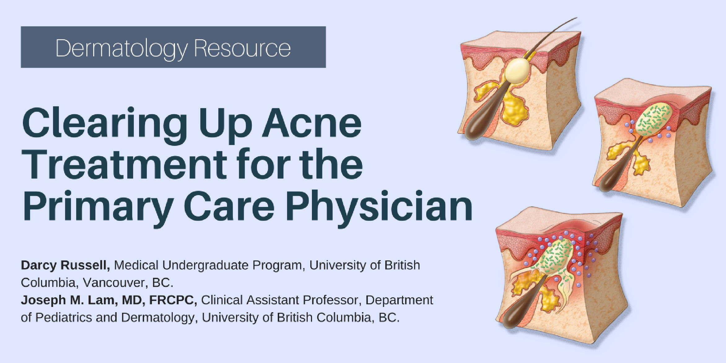
Dermatology

Disclaimer:  While every attempt is made to ensure that drug dosages provided within the text of this journal and the website are accurate, readers are urged to check drug package inserts before prescribing. Views and opinions in this publication and the website are not necessarily endorsed by or reflective of those of the publisher.

Briar Findlay1Joseph M. Lam, MD, FRCPC,2

1 Paediatric Resident, BC Children's Hospital, Vancouver, BC.
2Associate Clinical Professor, Department of Paediatrics, Associate Member, Department of Dermatology, University of British Columbia.

CLINICAL TOOLS

Abstract: Many dermatoses occur in the pediatric population that can mimic atopic dermatitis based on their morphology or their propensity for triggering itch. This review will highlight some of the common skin conditions that can mimic atopic dermatitis, their typical response to topical corticosteroids and helpful features that can help distinguish these conditions from atopic dermatitis.
Key Words: atopic dermatitis mimickers, topical corticosteroids, chronic inflammatory skin disease, paediatrics.
Many dermatitic eruptions can mimic atopic dermatitis but features such as their typical response to topical corticosteroids can be a helpful distinguishing feature.
Some atopic dermatitis mimickers can worsen with topical corticosteroids and these include periorificial dermatitis and tinea corporis.
Some atopic dermatitis mimickers will only partially improve with topical corticosteroids alone and these include allergic contact dermatitis and molluscum dermatitis.
Other atopic dermatitis mimickers such as psoriasis and seborrheic dermatitis can respond to topical corticosteroids and the correct diagnosis can be made using other morphological or historical features.
AD is a prevalent, chronic and relapsing condition in infancy and childhood.
Morphology, distribution and age of onset can be important in distinguishing between AD and common mimickers.
Response to corticosteroids is not diagnostic for AD as many mimickers may have an initial or complete response to topical corticosteroids; however, corticosteroid usage in some mimickers of AD may lead to complications and unnecessary side effects of topical corticosteroids.
To have access to full article that these tools were developed for, please subscribe. The cost to subscribe is $80 USD per year and you will gain full access to all the premium content on www.healthplexus.net, an educational portal, that hosts 1000s of clinical reviews, case studies, educational visual aids and more as well as within the mobile app.
Disclaimer at the end of each page

Linda Yang, BSc,1Joseph M. Lam, MD, FRCPC,2

1 Faculty of Medicine, University of British Columbia, Vancouver, BC.
2Clinical Associate Professor, Department of Pediatrics, Associate Member, Department of Dermatology and Skin Sciences, University of British Columbia, Vancouver, BC.

CLINICAL TOOLS

Abstract: Atopic dermatitis is a common pediatric disease with a chronic relapsing-remitting course, causing distress to patients and family. In patients who remain recalcitrant following treatment with topical steroids, adjunctive therapies including bleach baths, wet wraps and phototherapy as well as systemic immunosuppressants may be considered. Many novel therapies are in development and act on various aspects of the immunologic cascades involved in atopic dermatitis. The following review briefly summarizes up-to-date evidence for the use of these therapies in the pediatric population.
Key Words: atopic dermatitis, pediatric disease, therapies.
Topical corticosteroids, the first-line treatment for atopic dermatitis, can be optimized with usage of an appropriate amount and within a supportive, therapeutic alliance.
Those who fail to improve with topical corticosteroids may benefit from adjunctive treatment with wet wraps, bleach baths and phototherapy with narrowband UV therapy. These have been shown to be efficacious with a minimal side effect profile.
In those who remain recalcitrant, a brief course of immunosuppressants may be indicated. Methotrexate, azathioprine and cyclosporine have evidence in the pediatric population. Of these, methotrexate has been shown to have the most sustained duration of remission.
A recent explosion of novel immunomodulators and biologics may redefine atopic dermatitis treatment. Crisaborole is a topical PDE4 inhibitor, which has been approved for used in children. Dupilumab is an injectable monoclonal antibody, which has recently been approved for the adult population and remains off-label in pediatrics.
Monotherapy when possible and regular check-ins with parents can improve adherence to topical steroid regimens, particularly within the first 3 days of treatment.
The American Academy of Dermatology recommends the use of bleach baths (1/2 cup of 6% household bleach in a 150L bathtub full of water) for 5 to 10-minute intervals 2-3 times weekly as an adjunct to topicals.
To have access to full article that these tools were developed for, please subscribe. The cost to subscribe is $80 USD per year and you will gain full access to all the premium content on www.healthplexus.net, an educational portal, that hosts 1000s of clinical reviews, case studies, educational visual aids and more as well as within the mobile app.
Disclaimer at the end of each page

Miriam Armanious, BSc, MD candidate,1 Joseph M. Lam, MD, FRCPC,2

1Michael G. DeGroote School of Medicine, McMaster University, Hamilton, Ontario.
2Clinical Associate Professor, Department of Pediatrics, Associate Member, Department of Dermatology and Skin Sciences, University of British Columbia, Vancouver, BC.

CLINICAL TOOLS

Abstract: Superficial fungal infections are a common occurrence in adults and children alike. Dermatophytes are the primary cause of these infections, which generally present as erythematous, scaling, annular lesions. Also referred to as "tinea", these infections are classified based on where they are found on the body, as different locations can have slightly different presentations and treatment requirements. This article provides an overview of these various presentations of dermatophyte infections and their risk factors, as well as recommended therapies.
Key Words: dermatophytes, fungal infections, therapies.
Dermatophyte infections, also known as tinea, are very common fungal infections in humans. They occur on the superficial skin, hair, and nails, and can present in many different locations on the body.
Tinea captis is most common in children and can cause hair loss or abscess formation.
When tinea infections are treated with topical corticosteroids, they become harder to detect and are referred to as tinea incognito.
Tinea infections are common, but should be confirmed with KOH microscopy and/or culture from a skin scraping, nail clipping, or hair sample.
Tinea capitis can be mistaken for eczema or seborrheic dermatitis
Check patients who have tinea infection for tinea pedis, since this is a common source of infection for sites on the rest of the body
Treatment for dermatophyte infections can include oral antifungal agents such as terbinafine or grise-ofulvin in a weightdependent dose, or topical antifungal agents. Systemic agents are generally re-served for presentations that penetrate hair follicles and nails, or those that are refractory to topical treatment.
To have access to full article that these tools were developed for, please subscribe. The cost to subscribe is $80 USD per year and you will gain full access to all the premium content on www.healthplexus.net, an educational portal, that hosts 1000s of clinical reviews, case studies, educational visual aids and more as well as within the mobile app.
Disclaimer at the end of each page

Lauren Schock, BSc, MD Program,1 Joseph M. Lam, MD, FRCPC,2

1Cumming School of Medicine, University of Calgary, Calgary, AB.
2Clinical Associate Professor, Department of Pediatrics, Associate Member, Department of Dermatology and Skin Sciences, University of British Columbia, Vancouver, BC.

CLINICAL TOOLS

Abstract: Yellow-hued papules and plaques in children can be difficult to differentiate as many causes are rare and may not be frequently outside of specialty pediatric dermatology settings. We will review some of the common and concerning yellow-brown papules and plaques found in infants and children and discuss appearance and distribution, pathophysiology, associated findings, and management.
Key Words: dermatology, pediatric, yellow lesions.
Nevus sebaceous typically grow in proportion with patients in early childhood. Excision should be deferred until adolescence to avoid the use of general anesthetic and an informed decision can be made by the child.
Benign cephalic histiocytosis and juvenile xanthogranuloma are both forms of non-Langerhans cell histiocytosis and are benign and self limited.
Consider a diagnosis of tuberous sclerosis in any child presenting with connective tissue nevi, especially if white macules, angiofibroma, or periungual fibroma are also found.
Screen children with necrobiosis lipodica for retinopathy and neuropathy.
Use your hands – rub a suspected lesion of mastocytosis; if urticaria is elicited (a red, itchy, swollen papule or plaque), you have found Darier's sign. Mastocytosis is likely. Be prepared to treat the child with antihistamines if needed.
Juvenile xanthogranulomas are more common under two years of age, and typically appear on the head and neck. Cutaneous xanthomas often occur overlying tendons, or as grouped papules over the extensor surfaces and buttocks.
To have access to full article that these tools were developed for, please subscribe. The cost to subscribe is $80 USD per year and you will gain full access to all the premium content on www.healthplexus.net, an educational portal, that hosts 1000s of clinical reviews, case studies, educational visual aids and more as well as within the mobile app.
Disclaimer at the end of each page

Kailie Luan,1 Joseph M. Lam, MD, FRCPC,2

1Faculty of Medicine, University of Alberta, Edmonton, AB.
2Clinical Assistant Professor, Department of Pediatrics and Dermatology, University of British Columbia, BC.

CLINICAL TOOLS

Abstract: Alopecia areata is a chronic immune-mediated disorder that causes nonscarring hair loss. Although most commonly causing discrete hair loss on the scalp, the condition can affect any hair bearing area of the body and cause significant emotional and psychosocial distress. While intralesional glucocorticoids are often used as initial treatment for adults with the condition, therapeutic options for children are more limited with concerns of treatment tolerability and potential side effects. This article aims to provide an overview of alopecia areata with particular focus on managing this chronic condition in children.
Key Words: Alopecia areata, clinical presentation, diagnosis, management, pediatrics.
Alopecia areata is a chronic relapsing disorder characterized by non scarring hair loss that can affect any hair-bearing area of the body
While intralesional glucocorticoids are often used as initial treatment for adults, potent topical corticosteroids are effective as first line therapy in children due to better treatment tolerability
The diagnosis is generally made on clinical grounds with the majority of patients presenting with limited patchy disease affecting the scalp
In cases of inadequate response, topical minoxidil or immunotherapy are additional options, with systemic corticosteroids and immunosuppressive agents reserved for refractory cases, and IL-2 and JAK inhibitors as new emerging therapies for AA
Not all patients with alopecia areata require treatment as up to 50 percent of patients with limited alopecia areata will experience spontaneous regrowth of hair.4
Due to the benign nature of alopecia areata, and spontaneous remission is common, watchful waiting is considered a reasonable option in cases of limited disease.
To have access to full article that these tools were developed for, please subscribe. The cost to subscribe is $80 USD per year and you will gain full access to all the premium content on www.healthplexus.net, an educational portal, that hosts 1000s of clinical reviews, case studies, educational visual aids and more as well as within the mobile app.
Disclaimer at the end of each page

Clinical Images: Cercarial Dermatitis

Kerry Gardner, MD,1 Joseph M. Lam, MD, FRCPC,2

1Resident, Department of Dermatology and Skin Science, University of British Columbia, BC.
2Clinical Assistant Professor, Department of Pediatrics and Dermatology, University of British Columbia, BC.

CLINICAL TOOLS

Abstract: Cercarial dermatitis (swimmers' itch) consists of urticarial papules that form when the larvae of avian parasitic flatworms penetrate the skin. Cercarial dermatitis is common in the summer months where heaviest recreational swimming occurs, and when the temperature is ideal for amplified schistosome development.
Key Words: cercarial dermatitis (swimmers' itch), non-communicable, water-borne disease.
Cercarial dermatitis usually occurs with exposure to fresh water, but can occur with shallow salt water exposure as well.
The eruption typically occurs on uncovered skin 12-24 hours (up to 8 days) after exposure.
The eruption is self-limited, lasting 4-10 days (up to 20 days).
To have access to full article that these tools were developed for, please subscribe. The cost to subscribe is $80 USD per year and you will gain full access to all the premium content on www.healthplexus.net, an educational portal, that hosts 1000s of clinical reviews, case studies, educational visual aids and more as well as within the mobile app.
Disclaimer at the end of each page

Darcy Russell,1 Joseph M. Lam, MD, FRCPC,2

1Medical Undergraduate Program, University of British Columbia, Vancouver, BC.
2Clinical Assistant Professor, Department of Pediatrics and Dermatology, University of British Columbia, BC.

CLINICAL TOOLS

Abstract: Acne vulgaris is a common skin condition encountered in family practice and can cause significant distress during adolescence. Treatment options discussed include topical benzyl peroxide, topical retinoids, oral and topical antibiotics, hormonal therapy, and isotretinoin. The following review article provides up-to-date recommendations for treating mild to severe pediatric acne.
Key Words: acne vulgaris, adolescence, treatment, pathogenesis.
The differential diagnosis for acne in adolescence includes corticosteroid induced acne, folliculitis, keratosis pilaris, papular sarcoidosis, perioral dermatitis, pseudofolliculitis barbae, and tinea faceie.
Acne may be classified as mild, moderate or severe based on the number and type of lesions involved as well as the total surface area involved.
Acne therapy is targeted at treating as many pathogenic factors as possible.
Topical fixed-dose combination therapies can be used for all types and severities of acne in children 9 years of age and older.
Both topical and oral antibiotics work by inhibiting P acnes protein synthesis and decreasing inflammation.
Do not be afraid of isotretinoin. It can be used first line in patients with severe nodular and/or inflammatory acne, acne conglobata, and recalcitrant acne.12 It is the only treatment that targets all four pathogenic factors implicated in acne vulgaris and can permanently decrease acne.
To have access to full article that these tools were developed for, please subscribe. The cost to subscribe is $80 USD per year and you will gain full access to all the premium content on www.healthplexus.net, an educational portal, that hosts 1000s of clinical reviews, case studies, educational visual aids and more as well as within the mobile app.
Disclaimer at the end of each page

Rebeca Pinca, MD,1 Joseph M. Lam, MD, FRCPC,2

1Department of Dermatology & Skin Science, University of British Columbia, BC.
2Clinical Assistant Professor, Department of Pediatrics and Dermatology, University of British Columbia, BC.

CLINICAL TOOLS

Abstract: Dermatology is a visual specialty, yet palpation can also play an important diagnostic role. We present five dermatologic diagnoses that can be made at point of care by palpation or physical manoeuvres, potentially reducing unnecessary investigations, such as biopsies.
Key Words: Dermatofibromas, pilomatricomas, mastocytomas, spider angiomas, terra firma-forme dermatitis.
Dermatofibromas and pilomatricomas are benign papulonodular lesions that can be differentiated by the dimple sign, and the teeter-totter sign or tent sign, respectively.
Solitary mastocytomas can be diagnosed by Darier sign, whereby rubbing of the lesion causes a wheal and pruritus.
Spider angiomas can be diagnosed by diascopy, which involves the application of gentle downward pressure with a glass slide on the skin, resulting in blanching of the telangiectasia.
Terra firma-forme dermatitis is a benign discoloration that can be diagnosed, and treated, by gentle rubbing with isopropyl alcohol.
These dermatologic physical examination manoeuvres are quick, cost-effective, point-of-care diagnostic tools.
If in doubt, do not hesitate to biopsy lesions that appear suspicious.
To have access to full article that these tools were developed for, please subscribe. The cost to subscribe is $80 USD per year and you will gain full access to all the premium content on www.healthplexus.net, an educational portal, that hosts 1000s of clinical reviews, case studies, educational visual aids and more as well as within the mobile app.

Lisa M. Flegel,1 Joseph M. Lam, MD, FRCSC,2

1Medical Degree Undergraduate Program, Northern Medical Program, University of British Columbia, BC.
2Clinical Assistant Professor, Department of Pediatrics and Dermatology, University of British Columbia, BC.

CLINICAL TOOLS

Abstract: Hyperpigmented lesions are common in the pediatric population and identifying their etiologies can be challenging for physicians. Patients and caregivers may worry that hyperpigmented lesions are dangerous, associated with an internal illness or that they may lead to skin cancers. Having a better understanding of the causes and natural histories of these lesions may help to guide management and alleviate worry. This review article will provide an overview of select common and uncommon causes of hyperpigmented skin lesions in children.
Key Words: hyperpigmentation, pediatric.
1. Most hyperpigmented lesions in children do not require treatment aside from for cosmesis.
2. Features of malignant melanoma in children include: non-pigmented, uniform color, variable diameter, nodular lesions, and occurring de novo.
3. Parents and children should be warned that melanocytic nevi will grow as their child grows, but growth should be proportionate.
4. The risk of melanocytic nevi becoming malignant melanoma in children is very small.
In children with numerous melanocytic nevi, a good rule of thumb is to look for the 'ugly duckling' mole.
To track lesions over time, parents can develop a routine of taking a picture each year on the child's birthday.
To have access to full article that these tools were developed for, please subscribe. The cost to subscribe is $80 USD per year and you will gain full access to all the premium content on www.healthplexus.net, an educational portal, that hosts 1000s of clinical reviews, case studies, educational visual aids and more as well as within the mobile app.