Disclaimer: While every attempt is made to ensure that drug dosages provided within the text of this journal and the website are accurate, readers are urged to check drug package inserts before prescribing. Views and opinions in this publication and the website are not necessarily endorsed by or reflective of those of the publisher.
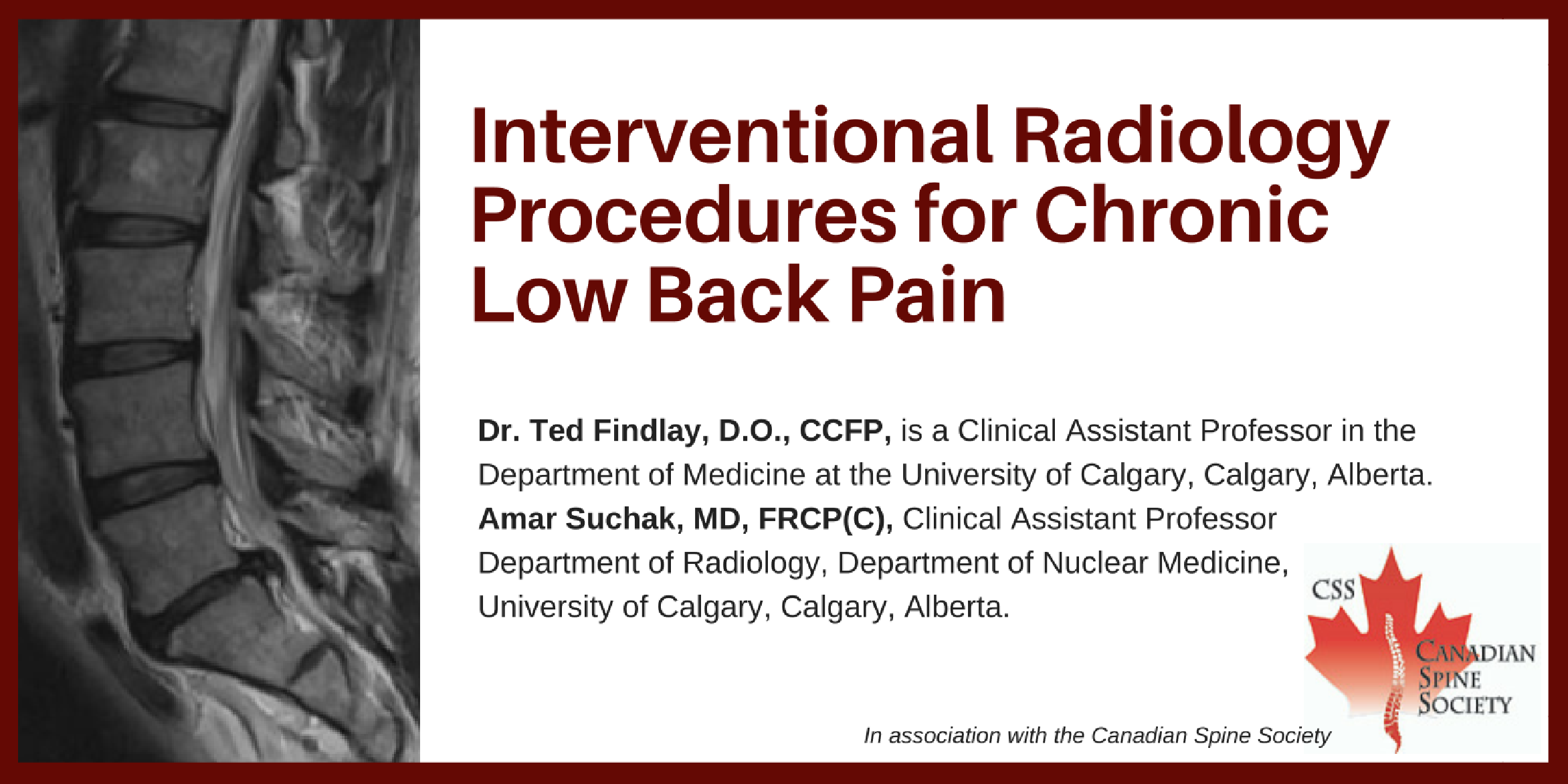
Dr. Ted Findlay, D.O., CCFP,1 Amar Suchak, MD, FRCP(C), 2
1is a Clinical Assistant Professor in the
Department of Medicine at the University of Calgary. He is
also in a Private Family Medicine practice. In addition he
is on Medical Staff at Alberta Health Services, Calgary
Zone in Calgary, Alberta.
2Clinical Assistant Professor Department of
Radiology, Department of Nuclear Medicine, University of
Calgary, Calgary, Alberta.
CLINICAL TOOLS
| Abstract: There is an increasing availability and clinical use of interventional radiological techniques for patients with low back pain. This can be a valuable additional tool in the management of low back pain that has not responded to conservative treatment. However, the clinical indications and appropriate uses as well as cautions that apply to this treatment modality are in many cases less well understood by the primary care practitioner. The objective of this article is to review clinical scenarios in which these procedures are commonly considered, as well as their limitations. The field of interventional radiology is one that is rapidly evolving and an area of active clinical research. It is important for the primary care practitioner to have a basic understanding of the current state of the art in order to have an informed discussion with their patients who may be seeking advice on this treatment option. |
| Key Words: Low back pain; treatment; interventional radiology definitions; interventional radiology indications; interventional radiology complications. |
Members of the College of Family Physicians of Canada may claim MAINPRO-M2 Credits for this unaccredited educational program.
www.cfpc.ca/Mainpro_M2You can take quizzes without subscribing; however, your results will not be stored. Subscribers will have access to their quiz results for future reference.


| 1. In patients carefully selected by clinical and radiological examination, there can be satisfying clinical gains from the use of currently available interventional radiologic procedures. |
| 2. One must not assume that abnormal findings on radiologic imaging immediately explains the anatomical cause of a patient's low back pain; a corresponding accurate history and physical examination is ideal prior to commencing injections. |
| 3. When successful, the gains from radiological interventions should be considered one portion of a broader clinical treatment plan, rather than the entire plan of management. |
| 4. Unsuccessful interventional procedures should not be repeated. |
| 1. Do not apply repeated interventional procedures with an expectation that one of them will find the target source of the patient's low back pain. |
| 2. Although they may be uncommon, interventional radiology risks can occur and the referring physician should be cognizant of these dangers that accumulate with repeated interventions. |
| To have access to full article that these tools were developed for, please subscribe. The cost to subscribe is $80 USD per year and you will gain full access to all the premium content on www.healthplexus.net, an educational portal, that hosts 1000s of clinical reviews, case studies, educational visual aids and more as well as within the mobile app. |
Dealing with Family Strife
Deck
Physicians usually become adept at choosing medications for the complaints and illnesses that patients bring to their attention.
Thumbnail Image


One always hopes that as medical practitioners, we will be able to focus our attention on the medical issues faced by seniors and help families cope with the fears, disappointments and tragedies that are faced by loved ones in the midst of what are often life-altering illnesses.
Topic
Section
- Read more about Dealing with Family Strife
- Log in or register to post comments
Editor's Note, Volume 6 Issue 4

D’Arcy Little, MD, CCFP, FRCPC
Medical Director, JCCC and HealthPlexus.NET

Beyond Medications for Dementia
Deck
Physicians usually become adept at choosing medications for the complaints and illnesses that patients bring to their attention.
Thumbnail Image


Physicians usually become adept at choosing medications for the complaints and illnesses that patients bring to their attention.
Topic
Section
- Read more about Beyond Medications for Dementia
- Log in or register to post comments
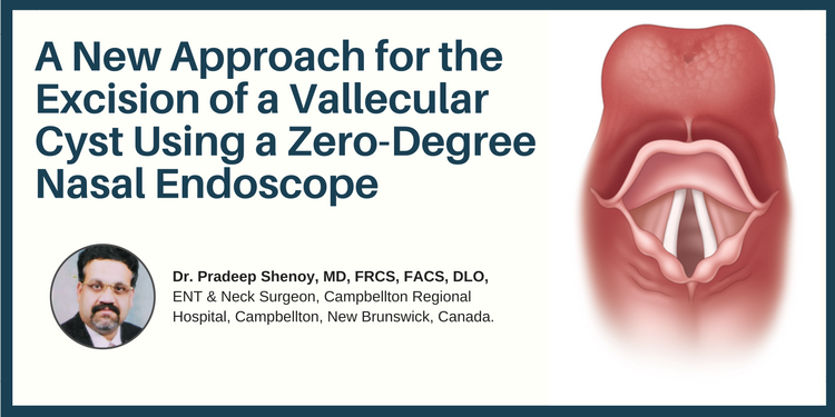
Dr. Pradeep Shenoy, MD, FRCS, FACS, DLO,
ENT & Neck Surgeon, Campbellton Regional Hospital, Campbellton, New Brunswick, Canada.
CLINICAL TOOLS
| Abstract: This case study reviews the clinical presentation and treatment of a patient’s vascular cyst. Though it is a rare diagnosis/condition, it could be a medical emergency for an individual of any age. Described here is a new approach for the complete excision of the vallecular cyst. |
| Key Words: vallecular cyst, excision, zero-degree nasal endoscope |

| Vallecular cysts are retention cysts in the Vallecular—a space between the base of the tongue, epiglottis and lateral pharyngeal wall. |
| They are triggered by acid reflux and smoking. |
| Vallecular cyst can be seen in CT Scan and laryngoscopy examination. |
| The access is difficult trans-orally. Here we are describing New approach using a zero-degree endoscope. |
| Vallecular cysts are rare in the paediatric and adult age groups. |
| Vallecular cysts can present as asymptomatic when small, however, when big they can present as a feeling of some food stuck in the throat or pain. |
| In emergency situation can block the food and airway passage and require emergency treatment. |
| To have access to full article that these tools were developed for, please subscribe. The cost to subscribe is $80 USD per year and you will gain full access to all the premium content on www.healthplexus.net, an educational portal, that hosts 1000s of clinical reviews, case studies, educational visual aids and more as well as within the mobile app. |
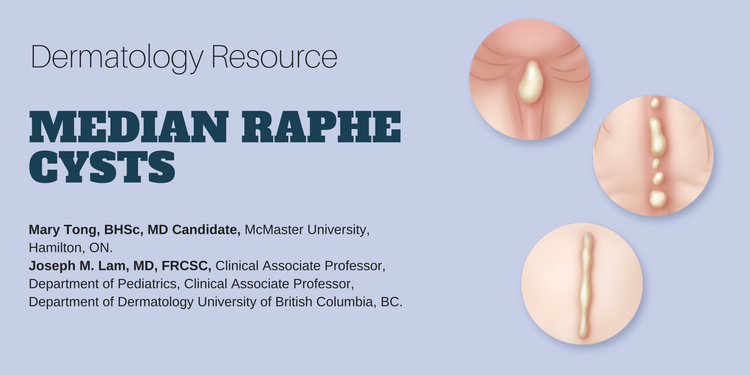
Mary Tong, BHSc, MD Candidate,1 Joseph M. Lam, MD, FRCSC,2
1McMaster University, Hamilton, ON.
2Clinical Associate Professor, Department of Pediatrics, Clinical Associate Professor, Department of Dermatology University of British Columbia, BC.
CLINICAL TOOLS
Abstract: Median raphe cysts are rare congenital lesions caused by a defect in embryological development of the male genitalia. They can present as solitary or multiple papules along the median raphe from urethral meatus to the anus. Although they are asymptomatic during childhood, they can cause problems later on as they increase in size. Surgical excision of the lesion is not necessary unless the patient becomes symptomatic.  |
| Key Words: median raphe cysts, congenital lesions, treatment, management. |


| Median raphe cysts are benign cysts that can be present at birth, or acquired due to trauma or infection in the genitalia area. |
| Histologically, the cysts can have pseudo stratified columnar, squamous cell, or glandular epithelium, or a mixture of these cells. |
| Although these cysts are asymptomatic during childhood, they should be monitored overtime because they may cause problems as they increase in size with time. |
| Because these are benign malformations, median raphe cysts do not require excision unless they cause problems such as pain, problems with urination or sexual activity, or for cosmetic reasons. |
| Median raphe cysts are benign lesions that may be caused be a defect in the embryological development of the male genitalia. |
| The differential diagnoses of median raphe cyst include glomus tumor, dermoid cyst, pilonidal cyst, epidermal inclusion cyst, urethral diverticulum, and steatocystoma. |
| Treatment for asymptomatic median raphe cyst is not necessary but surgical excision can be considered if the cyst is causing problems or for cosmetic reasons. |
| To have access to full article that these tools were developed for, please subscribe. The cost to subscribe is $80 USD per year and you will gain full access to all the premium content on www.healthplexus.net, an educational portal, that hosts 1000s of clinical reviews, case studies, educational visual aids and more as well as within the mobile app. |